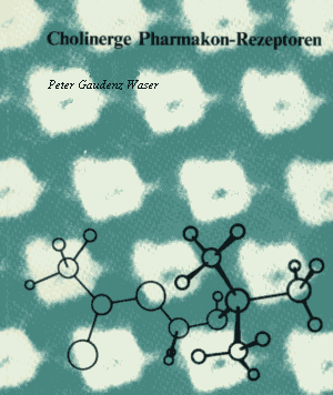
Figure on frontispiece:
Acetylcholine receptors in membrane fragments of electric organs (Torpedo californica). Electronmicrograph with high resolution, enhanced by phase averaging with computer.
when small coins or buttons (Ø 10 mm) are placed on the pores in this figure, a picture illustrating how the curare molecules (see fig. 21) sit on the postsynaptic membrane is produced. Magnification 5 000 000-fold
Contrast staining: Uranyl acetate (M.J. Ross et al., 1977)
Structure of Acetylcholine determined by X-ray diffraction (R. W. Baker et al., 1971).
Magnification: 100 000 000-fold. A 20-fold reduction of the structure of acetylcholine permits the direct comparison of molecule with the pictured receptor complex.
Acetylcholin-Rezeptoren in Membranfragmenten des elektrischen Organs des Zitterrochens (Torpedo californica). Elektronenmikroskopische Aufnahme mit hoher Auflösung, verstärkt durch Phasenmittelung mit Computer.
Wenn kleine Münzen oder Knöpfe (Ø 10 mm) auf die abgebildeten Poren gelegt werden, entsteht das Bild einer mit Curaremolekülen (siehe Bild 21) besetzten postsynaptischen Membran. Vergrösserung 5 000 000fach
Kontrastfärbung: Uranylacetat (M.J. Ross et al., 1977)
Struktur des Acetylcholins, bestimmt mit Röntgenbeugungsanalyse (R. W. Baker et al., 1971).
Vergrösserung: 100 000 000fach. Eine 20fache Verkleinerung der Acetylcholinstruktur erlaubt einen direkten Grössenvergleich zwischen Molekül und abgebildetem Rezeptorkomplex.
Zusammenfassung
Um die Jahrhundertwende wurden Pharmakon-Rezeptoren zum ersten Mal von J. N. Langley und P. Ehrlich durch ihre Versuche mit Nikotin, Curare und chemotherapeutischen Arsenverbindungen nachgewiesen und als Wirkungsorte in der Zelle beschrieben. Es wurde rasch erkannt, dass ihre Struktur und Verteilung in den Organen die spezifische Einwirkung Von Medikamenten ermöglicht. Nach dem Mechanismus von Schlüssel und Schloss wurden anfänglich vor allem die Schlüssel, d.h. chemische Verbindungen mit starker biologischer Wirkung, untersucht. Entscheidend für die Bindung an Rezeptoren oder Enzyme sind unter anderem die chemische Struktur, Grösse, Ladungsverteilung, Lipophilität, pharmakophore Gruppen und die Flexibilität des Moleküls. Eine Wirkung wird aber erst ausgelöst, wenn eine Reihe von meist wenig erkannten Veränderungen, häufig in der Zellmembran, abläuft:
Öffnung von Ionenkanälen, Freisetzung eines zweiten Boten wie Calcium, Koppelung an Membranenzyme zur Aktivierung des Zellstoffwechsels.
Das Wesen und Funktionieren des Schlosses, d.h. der Rezeptoren, die aktiviert werden müssen, wird erst durch die spezifische Markierung von Rezeptorstellen und die quantitative Untersuchung der Reaktionsabläufe (Reaktionspartner, Zeitkonstanten, Energiebilanz) erfassbar. Mit radioaktiv markierten Pharmaka und sogenannten Affinitätsmarken werden lose bis irreversible Bindungen an spezifischen Rezeptorstellen geschaffen, welche dann ihre Isolierung und Charakterisierung ermöglichen.
Der cholinerge Rezeptor wird vom Neurotransmittor Acetylcholin erregt. Die nikotinisch cholinergen Rezeptoren finden sich in den Endplatten der Skelettmuskulatur, in nervösen Ganglien und im Zentralnervensystem. Mit Hilfe der Bindung von a-Bungarotoxin und Cobratoxinen konnten diese Rezeptoren vor allem aus den elektrischen Organen von Zitterrochen und Aalen isoliert werden. Ihr Molekülgewicht, die Struktur der fünf Untereinheiten, der Aufbau aus Glykoproteinen und die Verbindung zu Ionenkanälen in den nervösen Membranen werden jetzt untersucht. Die muscarinisch cholinergen Rezeptoren kontrollieren vor allem die glatte Muskulatur der inneren Organe sowie neuronale Synapsen des Gehirns und Herzens. Sie können spezifisch radioaktiv markiert werden, doch ist ihre Isolierung und Reinigung noch nicht abgeschlossen. Subtypen des muscarinischen Rezeptors sind besonders wichtig zur Steuerung der Herz- und Gehirnfunktionen. Muscarinische Rezeptoren sind anscheinend für das Gedächtnis und das kognitive Denken notwendig. Rezeptoranomalien der cholinergen Synapsen sind bekannt bei Myasthenia gravis (Muskelschwäche), bei seniler Demenz, bei Morbus Parkinson und eventuell bei Epilepsie.